Hematology · USMLE Step 1
Anemia workup for USMLE: start with the MCV
Sort any anemia by MCV, then iron studies, reticulocyte count or B12 and folate markers. One flowchart, one iron-studies grid and practice questions.
There are dozens of causes of anemia, but questions almost always give you the two numbers that narrow them down: the MCV and, for normocytic anemia, the reticulocyte count. Start there and the list shrinks to a handful.
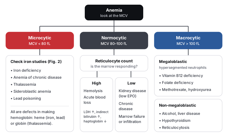
Microcytic: let the iron studies decide
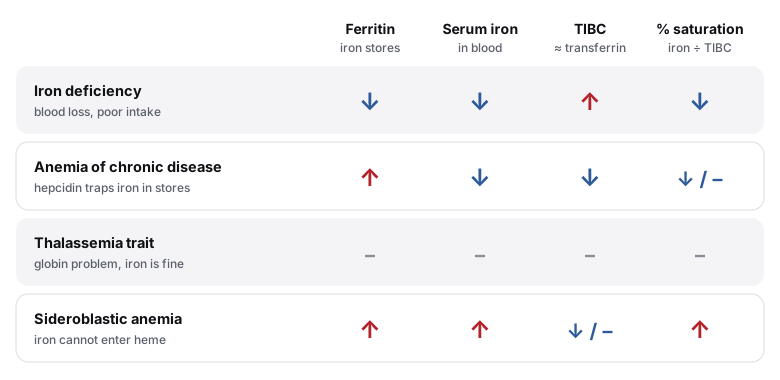
- Iron deficiency vs chronic disease: both have low serum iron. TIBC separates them: high when the body is hungry for iron, low when inflammation (hepcidin) locks it away.
- Thalassemia trait: very low MCV with a normal or high red cell count and normal iron studies. Confirm with hemoglobin electrophoresis (HbA₂ is raised in β-thalassemia trait).
- Sideroblastic anemia: ringed sideroblasts on the marrow iron stain. Causes include alcohol, lead, vitamin B6 deficiency (isoniazid) and copper deficiency.
Normocytic: is the marrow keeping up?
The raw reticulocyte percentage overstates the response when the hematocrit is low. Correct it: reticulocyte % × (Hct ÷ 45), then divide by a maturation factor (about 2 when the hematocrit is near 25%) to get the reticulocyte production index. Above 3 means the marrow is responding (hemolysis or blood loss); below 2 means it is not (underproduction).
| Intravascular | Extravascular | |
|---|---|---|
| Where cells are destroyed | Inside blood vessels | Macrophages in the spleen and liver |
| Both have | ↑ LDH, ↑ unconjugated bilirubin, ↓ haptoglobin, ↑ reticulocytes | |
| Extra clues | Hemoglobinuria, hemosiderinuria, schistocytes | Splenomegaly, spherocytes, pigment gallstones |
| Examples | Microangiopathic (TTP, HUS, DIC, mechanical valves), PNH, ABO transfusion reaction, G6PD crisis | Hereditary spherocytosis, warm autoimmune hemolysis, sickle cell disease |
| Disease | Clue | Confirm with |
|---|---|---|
| Hereditary spherocytosis | Spherocytes, ↑ MCHC, splenomegaly, family history | EMA binding test (flow cytometry) |
| G6PD deficiency | Hemolysis after sulfa drugs, primaquine, fava beans or infection; bite cells, Heinz bodies | G6PD activity, weeks after the episode |
| Autoimmune hemolysis | Warm (IgG): lupus, CLL, drugs. Cold (IgM): Mycoplasma, EBV | Direct Coombs (antiglobulin) test |
| PNH | Hemolysis with thrombosis (hepatic, cerebral veins), dark morning urine | Flow cytometry: CD55 and CD59 absent |
| Sickle cell disease | Vaso-occlusive crises, autosplenectomy, Howell-Jolly bodies | Hemoglobin electrophoresis (HbS) |
Smear clues
| Finding | Think of |
|---|---|
| Target cells | Thalassemia, HbC disease, liver disease, asplenia |
| Schistocytes | Microangiopathic hemolysis: TTP, HUS, DIC, mechanical valves |
| Spherocytes | Hereditary spherocytosis, warm autoimmune hemolysis |
| Bite cells, Heinz bodies | G6PD deficiency |
| Basophilic stippling | Lead poisoning, thalassemia |
| Howell-Jolly bodies | Asplenia or functional asplenia (sickle cell disease) |
| Teardrop cells | Myelofibrosis (marrow infiltration) |
| Hypersegmented neutrophils | B12 or folate deficiency |
Macrocytic: B12 or folate?
| Vitamin B12 deficiency | Folate deficiency | |
|---|---|---|
| Homocysteine | ↑ | ↑ |
| Methylmalonic acid | ↑ | Normal |
| Neurologic signs | Yes: subacute combined degeneration | No |
| Classic causes | Pernicious anemia, vegan diet, ileal resection or Crohn disease, metformin | Poor diet (alcohol use), pregnancy, methotrexate, phenytoin, trimethoprim |
Practice questions
Question 1HematologyThalassemia trait
A 23-year-old man has a hemoglobin of 11.6 g/dL found on routine testing. He feels well. MCV is 64 fL and the red cell count is 6.1 million/mm³. Ferritin and transferrin saturation are normal. Which test will most likely confirm the diagnosis?
A bone marrow biopsy is not needed for a stable, asymptomatic patient with microcytosis and normal iron studies. It is reserved for suspected marrow failure, infiltration, or ringed sideroblasts.
A very low MCV with a high red cell count and normal iron studies is the pattern of thalassemia trait. In iron deficiency the red cell count falls along with the hemoglobin. The Mentzer index (MCV ÷ RBC = 64 ÷ 6.1 ≈ 10.5) is below 13, which also favors thalassemia. Hemoglobin electrophoresis shows a raised HbA₂ in β-thalassemia trait (α-thalassemia trait can have a normal electrophoresis and needs genetic testing).
Lead poisoning causes a microcytic anemia with basophilic stippling, but it is expected in a child or a worker with exposure, abdominal pain or neuropathy, and the red cell count would be low, not high.
Occult gastrointestinal bleeding causes iron deficiency, but his ferritin and transferrin saturation are normal, so iron is not depleted.
B12 deficiency causes a macrocytic anemia with hypersegmented neutrophils. His MCV is 64 fL.
The answer is B.
Key takeaway
Marked microcytosis with a normal or high red cell count and normal iron studies is thalassemia trait: confirm with hemoglobin electrophoresis.
Thalassemia trait reduces globin chain production. The cells are small, but the marrow makes many of them, so the red cell count is high or normal (unlike iron deficiency, where it is low) and iron studies are normal. Patients are usually asymptomatic and need no treatment, but iron should not be given.
- Mentzer index (MCV ÷ RBC count) < 13: thalassemia. > 13: iron deficiency
- β-thalassemia trait: raised HbA₂ on electrophoresis; α-thalassemia trait may need genetic testing
- Smear: microcytes and target cells; ferritin and iron studies are normal
- Avoid iron supplements unless iron deficiency is proven
Question 2HematologyAnemia of chronic disease
A 62-year-old woman with long-standing rheumatoid arthritis has a hemoglobin of 10.2 g/dL and an MCV of 79 fL. Serum iron is low, TIBC is low and ferritin is high. Which molecule is most responsible for her anemia?
Erythropoietin falls in kidney disease, but the iron pattern here (low serum iron and TIBC with a high ferritin) is produced by iron being locked away, not by a lack of EPO. Inflammation does blunt the marrow's response to EPO, but hepcidin is the molecule that explains the iron findings.
This is anemia of chronic disease. Inflammatory cytokines, especially IL-6, increase hepcidin, which degrades ferroportin. Iron stays trapped in macrophages and is poorly absorbed from the gut: ferritin (stores) is high while serum iron and TIBC are low.
Haptoglobin binds free hemoglobin and is low in hemolysis. Hemolysis would raise LDH, indirect bilirubin and the reticulocyte count, not produce this iron pattern.
Intrinsic factor is needed for B12 absorption. Its absence (pernicious anemia) causes a macrocytic, megaloblastic anemia, not a normocytic or mildly microcytic one with a high ferritin.
The transferrin receptor is upregulated when cells need iron, as in iron deficiency. It is not the mediator of anemia of chronic disease.
The answer is B.
Key takeaway
In anemia of chronic disease, inflammation raises hepcidin: ferritin is high, serum iron and TIBC are low.
Hepcidin, made by the liver in response to IL-6, blocks iron export through ferroportin. Iron is trapped in storage and unavailable for hemoglobin, even though total body iron is normal or high. The anemia is usually normocytic, becoming mildly microcytic in long-standing disease. Treat the underlying disease; iron supplements do not help unless true iron deficiency coexists.
- Iron deficiency: ferritin low, TIBC high. Chronic disease: ferritin high, TIBC low
- Serum iron is low in both; TIBC separates them
- Seen in rheumatoid arthritis, chronic infection, cancer and chronic kidney disease
- Treat the cause; consider erythropoiesis-stimulating agents in selected patients (for example CKD)
Question 3HematologyVitamin B12 deficiency
A 66-year-old woman has fatigue and tingling in both feet. She has lost vibration sense in her toes. Hemoglobin is 9.1 g/dL, MCV 114 fL, and the smear shows hypersegmented neutrophils. Which pair of laboratory results is most likely?
Both would be normal in neither B12 nor folate deficiency. Normal values argue against a megaloblastic anemia, and this patient has megaloblastic features (MCV 114, hypersegmented neutrophils).
High homocysteine with a normal methylmalonic acid is the pattern of folate deficiency. Folate deficiency does not cause neuropathy, and this patient has loss of vibration sense.
B12 is a cofactor for two reactions: homocysteine → methionine (shared with folate) and methylmalonyl-CoA → succinyl-CoA (B12 only). B12 deficiency therefore raises both homocysteine and methylmalonic acid. The neuropathy and loss of vibration sense point to B12 rather than folate. Pernicious anemia (antibodies against intrinsic factor or parietal cells) is the most common cause at her age.
Normal homocysteine with a raised methylmalonic acid is unusual. In B12 deficiency homocysteine is also raised, because B12 is needed for the methionine synthase reaction too.
The answer is C.
Key takeaway
B12 deficiency raises both homocysteine and methylmalonic acid and causes neurologic signs; folate deficiency raises only homocysteine.
Megaloblastic anemia with hypersegmented neutrophils points to B12 or folate deficiency. Methylmalonic acid separates them: it rises only in B12 deficiency. Neuropathy, loss of vibration and position sense, or subacute combined degeneration make B12 the answer. Giving folate alone can correct the anemia while the nerve damage progresses.
- Megaloblastic anemia with hypersegmented neutrophils: B12 or folate deficiency
- Neuropathy and loss of vibration or position sense: B12, not folate
- B12 causes: pernicious anemia, vegan diet, ileal disease or resection, metformin
- Treat with B12 (intramuscular or high-dose oral); check B12 before giving folate
Go deeper in the Knowledge Hub: iron deficiency anemia, thalassemia, hemolytic anemia and vitamin B12 deficiency.