Pharmacology · USMLE Step 1
Antiarrhythmic drugs for USMLE: classes, mechanisms, ECG changes and side effects
Vaughan Williams classes I to IV explained with action potential diagrams: how each antiarrhythmic works, its ECG changes, uses and high-yield toxicities, plus USMLE-style questions.
Antiarrhythmics are a favorite Step 1 topic because one question can test three things at once: the ion channel a drug blocks, what that does to the ECG, and the side effect that gives the drug away. Learn them as a list of names and they blur together. Learn them from the action potential up and most questions answer themselves.
This guide covers the Vaughan Williams classes I to IV, the drugs outside the classes that still show up on exams (adenosine, magnesium, digoxin, ivabradine), and the clinical traps that turn up again and again in questions. There are three diagrams, a one-page summary table, and five practice questions at the end.
The two action potentials you need to know
Every antiarrhythmic acts on one or both of two kinds of cardiac action potential. Know which ion carries each phase and you can predict what a drug will do.
1. Fast-response cells: ventricular and atrial muscle, Purkinje fibers
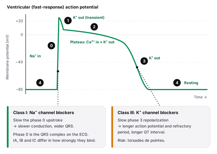
These cells rest at about −85 mV. Phase 0 is a rapid upstroke as voltage-gated Na⁺ channels open. Phase 1 is a brief repolarization from a transient K⁺ outflow. Phase 2 is the plateau, where Ca²⁺ entering through L-type channels balances K⁺ leaving. Phase 3 is repolarization as K⁺ channels carry the membrane back to rest, and phase 4 is the stable resting potential.
On the ECG, phase 0 across the ventricles corresponds to the QRS complex, and the end of phase 3 to the end of the T wave. So a drug that slows phase 0 widens the QRS, and a drug that delays phase 3 lengthens the QT.
2. Slow-response cells: the SA and AV nodes
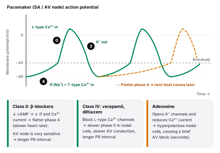
Nodal cells have no stable resting potential. During phase 4 they depolarize slowly on their own, driven by the "funny" current (If, carried mainly by Na⁺) and later T-type Ca²⁺ channels. When they reach threshold, phase 0 is a slower upstroke carried by L-type Ca²⁺ channels, not Na⁺. Phase 3 is K⁺-driven repolarization.
The slope of phase 4 sets the heart rate: steeper means faster. Sympathetic stimulation steepens it through β₁ receptors and cAMP; vagal tone and β-blockers flatten it. Conduction through the AV node depends on the Ca²⁺-driven upstroke, which is why calcium channel blockers and β-blockers both lengthen the PR interval.
Vaughan Williams classification at a glance
Use this table for revision once you have read the sections below.
| Class | Blocks | Drugs | ECG | Main uses | High-yield toxicity |
|---|---|---|---|---|---|
| IA | Na⁺ (moderate) + K⁺ | Quinidine, procainamide, disopyramide | ↑ QRS, ↑ QT | Atrial and ventricular arrhythmias; procainamide for stable pre-excited AF (WPW) | Torsades; quinidine: cinchonism; procainamide: drug-induced lupus; disopyramide: heart failure, anticholinergic effects |
| IB | Na⁺ (weak, fast dissociation) | Lidocaine, mexiletine | Little change | Ventricular arrhythmias, especially with ischemia | CNS effects (paresthesias, tremor, confusion, seizures); cardiovascular depression |
| IC | Na⁺ (strong, slow dissociation) | Flecainide, propafenone | ↑↑ QRS | Supraventricular arrhythmias, including AF, in structurally normal hearts | Proarrhythmia; increased mortality after MI (CAST) |
| II | β-adrenergic receptors | Metoprolol, esmolol, propranolol, atenolol | ↑ PR | Rate control in AF and flutter, SVT, after MI | Bradycardia, AV block, bronchospasm (nonselective), masked hypoglycemia; overdose → glucagon |
| III | K⁺ | Amiodarone, sotalol, dofetilide, ibutilide (and dronedarone) | ↑ QT | AF and flutter, ventricular tachycardia; amiodarone in cardiac arrest from VF or pulseless VT | Torsades (sotalol, dofetilide, ibutilide); amiodarone: lungs, liver, thyroid, skin, eyes |
| IV | L-type Ca²⁺ | Verapamil, diltiazem | ↑ PR | SVT, rate control in AF and flutter | Bradycardia, AV block, worsened heart failure; verapamil: constipation |
Class I: sodium channel blockers
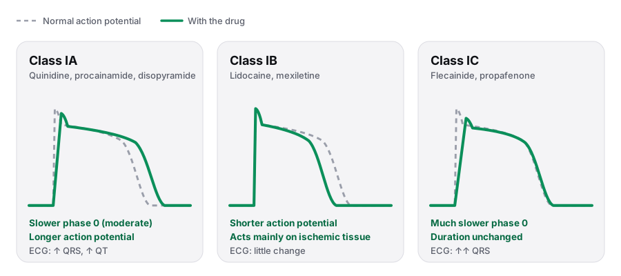
All Class I drugs block the fast Na⁺ channels that drive phase 0, so they slow conduction in atrial, ventricular and Purkinje tissue. They are use-dependent: they bind channels that are open or inactivated, so their effect grows at faster heart rates, when channels spend more time in those states. The three subclasses differ in how tightly they bind and in what else they block.
Class IA: quinidine, procainamide, disopyramide
Moderate Na⁺ block slows phase 0, and these drugs also block K⁺ channels, so the action potential and effective refractory period get longer. On the ECG: a wider QRS and a longer QT. They treat both atrial and ventricular arrhythmias.
- Procainamide is the classic choice for hemodynamically stable atrial fibrillation with an accessory pathway (Wolff-Parkinson-White). It causes a drug-induced lupus with antihistone antibodies, more often in slow acetylators. Its metabolite N-acetylprocainamide (NAPA) has Class III activity.
- Quinidine causes cinchonism (headache, tinnitus, hearing loss), diarrhea and thrombocytopenia, and can raise digoxin levels.
- Disopyramide has strong anticholinergic effects (urinary retention, dry mouth) and is negatively inotropic, so it can precipitate heart failure.
- All three prolong the QT and can cause torsades de pointes.
Class IB: lidocaine, mexiletine
Weak Na⁺ block with fast dissociation, so normal tissue at normal rates is hardly affected. IB drugs bind preferentially to inactivated channels, which are plentiful in depolarized, ischemic tissue. That makes them a fit for ventricular arrhythmias in the setting of ischemia. They shorten the action potential and change the ECG very little. Lidocaine is given IV; mexiletine is its oral relative.
Toxicity is mostly neurologic: perioral numbness, tremor, confusion and, at high levels, seizures. Like all antiarrhythmics they can depress cardiac function.
Class IC: flecainide, propafenone
Strong Na⁺ block with slow dissociation produces the most marked slowing of conduction and the widest QRS, with little change in action potential duration. They are used for supraventricular arrhythmias, including atrial fibrillation, but only in patients with a structurally normal heart.
Class II: beta blockers
Blocking β₁ receptors lowers cAMP in nodal cells, which reduces the If and Ca²⁺ currents. Phase 4 flattens (slower sinus rate) and conduction through the AV node slows (longer PR interval). Common examples are metoprolol (β₁-selective), esmolol (IV, very short-acting, easy to titrate), propranolol (nonselective) and atenolol.
Uses: ventricular rate control in atrial fibrillation and flutter, prevention of recurrent SVT, and arrhythmia prevention after myocardial infarction, where β-blockers also reduce mortality.
- Bradycardia, AV block and worsening of acute decompensated heart failure.
- Bronchospasm with nonselective agents; caution in asthma.
- They can mask the adrenergic warning signs of hypoglycemia (tremor, palpitations) in diabetes.
- Fatigue and erectile dysfunction.
- Overdose: bradycardia and hypotension; glucagon is the classic antidote because it raises cAMP without using β receptors.
Class III: potassium channel blockers
Blocking the K⁺ channels of phase 3 delays repolarization, so the action potential and effective refractory period lengthen and the QT interval grows. The drugs to know are amiodarone, sotalol, dofetilide and ibutilide.
- Sotalol is also a nonselective β-blocker, so it adds bradycardia and β-blocker side effects to the torsades risk.
- Ibutilide (IV) is used for rapid conversion of recent-onset atrial fibrillation or flutter; dofetilide (oral) keeps patients in sinus rhythm. Both carry a significant torsades risk and are started with ECG monitoring.
Amiodarone: the drug with the most side effects to know
Amiodarone mainly blocks K⁺ channels but also has Class I, II and IV effects. Despite prolonging the QT, it rarely causes torsades. It is used for atrial fibrillation and flutter, ventricular tachycardia, and in cardiac arrest from ventricular fibrillation or pulseless ventricular tachycardia. Its half-life is very long (weeks), so toxicity can persist after the drug is stopped.
| Organ | Toxicity | Monitoring |
|---|---|---|
| Lungs | Pulmonary fibrosis (interstitial pneumonitis): cough, dyspnea | Chest x-ray and pulmonary function tests |
| Thyroid | Hypothyroidism or hyperthyroidism: the molecule is about one-third iodine by weight, and it inhibits peripheral T4 to T3 conversion | Thyroid function tests |
| Liver | Raised transaminases, hepatotoxicity | Liver function tests |
| Skin | Photosensitivity; blue-gray skin discoloration | Examination; sun protection |
| Eyes | Corneal microdeposits; rarely optic neuropathy | Eye examination |
| Heart | Bradycardia, heart block | ECG |
| Interactions | Inhibits cytochrome P450 enzymes and P-glycoprotein: raises warfarin and digoxin levels | INR and digoxin levels |
For more depth, read the Knowledge Hub article on amiodarone and the one on long QT syndrome.
Class IV: calcium channel blockers
Verapamil and diltiazem, the non-dihydropyridine calcium channel blockers, block L-type Ca²⁺ channels in the SA and AV nodes. The nodal upstroke slows, AV conduction slows and its refractory period lengthens, so the PR interval increases. They are used to terminate and prevent re-entrant SVT and for ventricular rate control in atrial fibrillation and flutter.
- Adverse effects: bradycardia, AV block, hypotension and worsened heart failure (negative inotropy; avoid in heart failure with reduced ejection fraction). Verapamil characteristically causes constipation; peripheral edema and flushing are also seen.
- Both raise digoxin levels.
See the Knowledge Hub on calcium channel blockers and beta blockers for the full pharmacology.
Drugs outside the four classes
Adenosine
Adenosine acts on A1 receptors in the AV node: it increases K⁺ outflow (hyperpolarizing the cells) and reduces Ca²⁺ current, causing a transient AV block. It is the drug of choice to diagnose and terminate regular narrow-complex re-entrant SVT, such as AV nodal re-entrant tachycardia, after vagal maneuvers fail. Its half-life is under 10 seconds, so it is given as a rapid IV push followed by a saline flush.
- Side effects are brief but unpleasant: flushing, chest discomfort, dyspnea or bronchospasm, hypotension and a sense of impending doom.
- Theophylline and caffeine block adenosine receptors and blunt its effect; dipyridamole blocks adenosine reuptake and enhances it.
Magnesium
IV magnesium sulfate is first-line for torsades de pointes, even when the serum magnesium is normal, and it is also used in digoxin toxicity.
Digoxin
Digoxin inhibits the Na⁺/K⁺-ATPase, which raises intracellular Ca²⁺ (positive inotropy), and increases vagal tone, which slows AV conduction. It is used for rate control in atrial fibrillation, especially with heart failure.
- Toxicity: nausea, vomiting, blurred or yellow-tinted vision, and arrhythmias; hyperkalemia in acute overdose.
- Toxicity is more likely with renal failure, hypokalemia, and drugs that raise digoxin levels (verapamil, amiodarone, quinidine).
- Treatment: digoxin immune Fab (antibody fragments), correction of potassium, and magnesium for arrhythmias.
Ivabradine
Ivabradine blocks the If (funny) current in the SA node, flattening phase 4 and lowering the heart rate without affecting contractility. It is used in heart failure with reduced ejection fraction when the resting heart rate stays high despite a β-blocker. A distinctive side effect is luminous visual phenomena (phosphenes); it can also cause bradycardia.
Classic exam traps
- Atrial fibrillation with WPW. Avoid AV nodal blockers (adenosine, β-blockers, verapamil, diltiazem, digoxin): blocking the AV node can push conduction down the accessory pathway and trigger ventricular fibrillation. Stable patients get procainamide; unstable patients get synchronized cardioversion.
- A wide, twisting polymorphic VT with a long QT is torsades de pointes. Give IV magnesium; defibrillate if the patient is pulseless. Stop QT-prolonging drugs (Class IA and III, and many non-cardiac drugs) and correct low potassium and magnesium.
- Lupus-like illness with antihistone antibodies in a patient on an antiarrhythmic: procainamide.
- Pulmonary fibrosis plus thyroid and liver abnormalities: amiodarone.
- Tinnitus, headache and diarrhea: quinidine (cinchonism).
- Seizures or perioral numbness after an IV antiarrhythmic for VT: lidocaine.
- Adenosine "didn't work" in a heavy coffee drinker or a patient taking theophylline: receptor blockade blunts its effect.
- Prior MI and a prescription for flecainide: the answer is that it increases mortality.
To see these drugs in clinical context, read the Knowledge Hub articles on atrial fibrillation, supraventricular tachycardia, ventricular tachycardia and management of tachycardia.
Practice questions
Try each question before opening the answer.
Question 1PharmacologyAmiodarone toxicity
A 67-year-old man has had paroxysmal atrial fibrillation for 3 years, treated with a rhythm-control drug. Over the past 4 months he has developed increasing exertional dyspnea and a dry cough, and he has noticed a blue-gray discoloration of his face and hands. A chest x-ray shows bilateral reticular opacities. His serum TSH is elevated. Which drug is most likely responsible?
Pulmonary fibrosis (cough, dyspnea, reticular opacities), blue-gray skin discoloration and thyroid dysfunction together are classic amiodarone toxicity. The drug is about one-third iodine by weight, so it can cause hypothyroidism (as here) or hyperthyroidism, and its very long half-life means toxicity can persist for weeks after it is stopped.
Digoxin toxicity causes nausea and vomiting, yellow-tinted vision, confusion and arrhythmias. It does not cause pulmonary fibrosis, skin discoloration or thyroid disease.
Diltiazem, a non-dihydropyridine calcium channel blocker, causes bradycardia, AV block, hypotension and edema. It does not cause fibrosis or thyroid dysfunction.
Flecainide (Class IC) can be proarrhythmic, especially after a myocardial infarction (the CAST trial), but it is not associated with pulmonary fibrosis, skin discoloration or thyroid disease.
Metoprolol causes bradycardia, fatigue and can mask the symptoms of hypoglycemia. It does not cause pulmonary fibrosis or thyroid dysfunction.
The answer is A.
Key takeaway
Pulmonary fibrosis with thyroid dysfunction and blue-gray skin in a patient on an antiarrhythmic is amiodarone toxicity.
Amiodarone is a Class III drug that also has Class I, II and IV effects. It is effective but toxic to many organs because it is lipophilic, accumulates in tissues and has a half-life of weeks. Baseline and periodic monitoring of the lungs, thyroid, liver and eyes is standard.
- Toxicity: lungs (fibrosis), thyroid (hypo or hyper), liver, skin (photosensitivity, blue-gray), eyes (corneal deposits), heart (bradycardia, QT prolongation)
- Monitor TSH, liver tests, chest x-ray or PFTs, and ECG
- Inhibits CYP450 and P-glycoprotein: raises warfarin and digoxin levels
- Very long half-life: toxicity persists for weeks after stopping
Question 2PharmacologySupraventricular tachycardia
A 26-year-old woman has had palpitations for 30 minutes. Her blood pressure is 118/76 mm Hg and her pulse is 190/min and regular. An ECG shows a regular narrow-complex tachycardia with no visible P waves. Carotid sinus massage does not change the rhythm. Which drug is the most appropriate next step?
A stable, regular narrow-complex tachycardia that does not respond to vagal maneuvers is most likely AV nodal re-entrant tachycardia. A rapid IV push of adenosine hyperpolarizes AV nodal cells and causes a brief AV block that terminates the re-entry circuit. Its half-life is under 10 seconds, so warn the patient about flushing, chest discomfort and a sense of impending doom.
Amiodarone is not first-line for terminating re-entrant SVT. It acts much more slowly and has significant toxicity.
Digoxin acts too slowly and is used for rate control in atrial fibrillation, not for rapid termination of re-entrant SVT.
Lidocaine (Class IB) acts mainly on ischemic ventricular tissue. It has no role in a supraventricular re-entrant rhythm.
Procainamide is used for stable pre-excited atrial fibrillation (WPW) and some wide-complex tachycardias. It is not first-line for AV nodal re-entry and can cause hypotension.
The answer is A.
Key takeaway
Stable regular narrow-complex SVT that fails vagal maneuvers is terminated with IV adenosine (rapid push).
Most regular narrow-complex tachycardias are re-entrant circuits that run through the AV node. Vagal maneuvers come first. Adenosine produces a few seconds of AV block, which breaks the circuit. An unstable patient (hypotension, chest pain, altered mental status) gets synchronized cardioversion instead.
- Stable regular narrow-complex SVT: vagal maneuvers, then adenosine
- Unstable (hypotension, chest pain, altered mental status): synchronized cardioversion
- Caffeine and theophylline block adenosine; dipyridamole enhances it
- Can cause bronchospasm: use caution in asthma
Question 3PharmacologyTorsades de pointes
A 70-year-old woman was started on sotalol for atrial fibrillation 2 days ago. She has an episode of syncope while on telemetry. The monitor shows a polymorphic ventricular tachycardia whose QRS complexes twist around the baseline, preceded by a QTc of 560 ms. The rhythm stops on its own and she now has a pulse and a normal blood pressure. Which drug should be given now?
Amiodarone also prolongs the QT interval. It rarely causes torsades itself, but it is not the treatment for torsades from a long QT.
This is torsades de pointes caused by a QT-prolonging Class III drug. IV magnesium sulfate is first-line, even when the serum magnesium is normal. Sotalol should be stopped, and potassium and magnesium kept in the normal range. A pulseless patient needs defibrillation.
Procainamide (Class IA) prolongs the QT interval and can itself cause torsades. Giving it would make this worse.
Verapamil is used for supraventricular arrhythmias and can cause hypotension and hemodynamic collapse in a ventricular tachyarrhythmia.
The answer is B.
Key takeaway
Torsades de pointes (polymorphic VT with a long QT) is treated with IV magnesium sulfate, even with a normal magnesium level.
Drugs that block potassium channels lengthen repolarization and the QT interval, which can trigger early afterdepolarizations and torsades. Stop the offending drug, correct low potassium and magnesium, and give IV magnesium. If the patient is pulseless, defibrillate.
- Torsades: polymorphic VT with a long QT interval
- Causes: Class IA and III drugs, macrolides, antipsychotics, ondansetron, hypokalemia, hypomagnesemia, congenital long QT
- Treatment: IV magnesium sulfate; defibrillation if pulseless
- Stop QT-prolonging drugs and correct K⁺ and Mg²⁺; consider pacing or isoproterenol if it keeps recurring
Keep going
Related reading in the free Knowledge Hub: