Respiratory · USMLE Step 1
Hypoxemia and the A–a gradient: five causes, one flowchart
Sort every cause of hypoxemia with two questions: is the A–a gradient normal, and does oxygen fix it? Flowchart, V/Q diagram and practice questions.
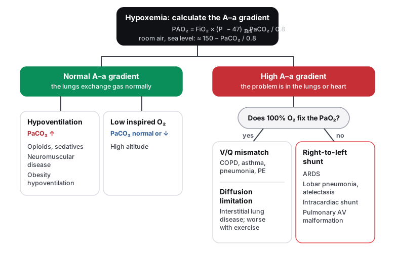
Every cause of a low PaO₂ falls into one of five groups, and two questions sort them: is the A–a gradient normal? and, if not, does extra oxygen fix it?
The A–a gradient in one line
It is the difference between the oxygen in the alveoli (PAO₂, calculated) and in arterial blood (PaO₂, measured). If the lungs pass oxygen across normally, the gap is small: roughly age ÷ 4 + 4 mm Hg, so under about 15 in young adults. A normal gap with a low PaO₂ means too little oxygen is reaching the alveoli; a wide gap means oxygen is not crossing into the blood.
Worked example: room air at sea level, PaCO₂ 40, PaO₂ 60. PAO₂ = 0.21 × (760 − 47) − 40 / 0.8 ≈ 150 − 50 = 100 mm Hg. The gradient is 100 − 60 = 40 mm Hg: wide, so the problem is in the lungs.
| Cause | PaCO₂ | A–a gradient | Corrects with O₂? | Examples |
|---|---|---|---|---|
| Hypoventilation | ↑ | Normal | Yes | Opioids, neuromuscular disease, obesity hypoventilation |
| Low inspired O₂ | ↓ (hyperventilation) | Normal | Yes | High altitude |
| V/Q mismatch | Normal or ↓ | ↑ | Yes | COPD, asthma, pneumonia, pulmonary embolism |
| Diffusion limitation | Normal or ↓ | ↑ | Yes | Interstitial lung disease, emphysema |
| Shunt | Normal or ↓ | ↑ | Little or not at all | ARDS, atelectasis, intracardiac shunt, pulmonary AV malformation |
DLCO measures diffusion. It falls in interstitial lung disease, emphysema, pulmonary vascular disease and anemia, and rises with polycythemia, alveolar hemorrhage and often asthma. It is normal in chronic bronchitis.
Why shunt is the one that oxygen cannot fix
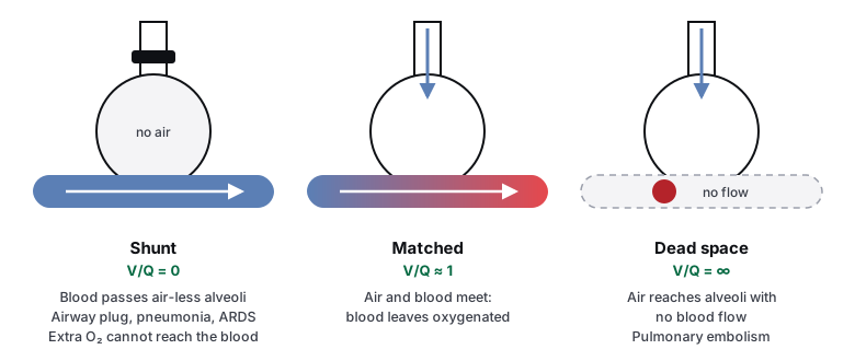
In V/Q mismatch, poorly ventilated units still get some air, so extra oxygen raises their alveolar PO₂ and the PaO₂ improves. In a shunt, the blood bypasses ventilated alveoli entirely. In the upright lung, the apex has the highest V/Q and the base the lowest.
The lung protects itself with hypoxic vasoconstriction: pulmonary arterioles constrict around poorly ventilated alveoli, diverting blood to better units (the opposite of systemic vessels). Chronic, widespread hypoxia (COPD, altitude) makes it global, causing pulmonary hypertension and right heart strain.
Low oxygen delivery with a normal PaO₂
PaO₂ only measures dissolved oxygen. Most oxygen rides on hemoglobin, so these conditions starve tissues while the PaO₂ looks fine.
| Condition | Pulse oximetry | O₂ content | Clues and treatment |
|---|---|---|---|
| Anemia | Normal | ↓ | Low hemoglobin; SaO₂ is normal |
| Carbon monoxide | Falsely normal | ↓ | Headache, fire or faulty heater; carboxyhemoglobin level; 100% or hyperbaric O₂ |
| Methemoglobinemia | About 85%, does not improve with O₂ | ↓ | Chocolate-brown blood after dapsone, nitrites or topical anesthetics (benzocaine); methylene blue |
| Cyanide | Normal | Normal | Cells cannot use O₂ (complex IV blocked): lactic acidosis, bright red venous blood; hydroxocobalamin |
Acclimatization to altitude
- Immediately: hyperventilation (peripheral chemoreceptors) → respiratory alkalosis.
- Over days: the kidneys excrete bicarbonate; 2,3-BPG rises (right-shifting the oxygen–hemoglobin curve); erythropoietin raises the red cell mass.
- Acetazolamide speeds acclimatization by causing a metabolic acidosis that drives ventilation.
Practice questions
Question 1RespiratoryHypoventilation
A 27-year-old man is found unresponsive after injecting heroin. His respirations are 5/min. Arterial blood gas on room air at sea level: PaO₂ 55 mm Hg, PaCO₂ 70 mm Hg. What is the cause of his hypoxemia?
Diffusion limitation (interstitial lung disease, emphysema) widens the A–a gradient. His gradient is normal, and diffusion limitation would not explain the raised PaCO₂.
PAO₂ ≈ 150 − 70 / 0.8 ≈ 62 mm Hg, so the A–a gradient is about 62 − 55 = 7 mm Hg: normal. A normal gradient with a high PaCO₂ means the lungs exchange gas normally but are not ventilated enough: hypoventilation. Opioids depress the respiratory drive in the medulla.
A shunt widens the A–a gradient and does not correct with 100% oxygen. His gradient is normal, and a shunt alone does not raise the PaCO₂, because CO₂ is so diffusible that the rest of the lung compensates.
V/Q mismatch also widens the A–a gradient, and he has no lung disease. A normal gradient and a high PaCO₂ point to inadequate ventilation of otherwise normal lungs.
Low inspired oxygen (altitude) lowers the PaO₂ with a normal gradient, but the PaCO₂ would be low from compensatory hyperventilation, not raised. He is at sea level on room air.
The answer is B.
Key takeaway
A low PaO₂ with a high PaCO₂ and a normal A–a gradient is hypoventilation.
Hypoxemia is sorted first by the A–a gradient. A normal gradient means the lungs transfer oxygen normally, so the cause is too little oxygen reaching the alveoli: hypoventilation (PaCO₂ raised) or low inspired oxygen (PaCO₂ low). Opioid overdose is the classic hypoventilation, and naloxone reverses it.
- PAO₂ = FiO₂ × (Patm − 47) − PaCO₂ / 0.8; on room air at sea level ≈ 150 − PaCO₂ / 0.8
- Normal A–a gradient: roughly age ÷ 4 + 4 (under about 15 mm Hg in young adults)
- Normal gradient: hypoventilation or low inspired oxygen
- Causes of hypoventilation: opioids, sedatives, neuromuscular disease, obesity hypoventilation
Question 2RespiratoryRight-to-left shunt
A 52-year-old woman with severe pancreatitis develops acute respiratory failure with bilateral infiltrates on chest x-ray. Her PaO₂ is 58 mm Hg while she breathes 100% oxygen. Which mechanism best explains her hypoxemia?
Diffusion limitation responds well to supplemental oxygen, because a higher alveolar PO₂ drives more oxygen across a thickened barrier. Her PaO₂ barely improves on 100% oxygen.
Hypoventilation corrects with oxygen and gives a normal A–a gradient with a high PaCO₂. She is on 100% oxygen yet still severely hypoxemic, so the problem is not simply too little ventilation.
She is breathing 100% oxygen, so inspired oxygen is not low.
This is ARDS. Alveoli fill with protein-rich fluid and collapse, so blood flows past units that receive no air: an intrapulmonary shunt. That is why the PaO₂ stays low even on 100% oxygen: blood that never meets ventilated alveoli cannot pick up the extra oxygen.
V/Q mismatch improves with supplemental oxygen, because poorly ventilated alveoli still receive some air and a higher inspired oxygen raises their alveolar PO₂. A PaO₂ of 58 on 100% oxygen means blood is bypassing ventilated alveoli altogether.
The answer is D.
Key takeaway
Hypoxemia that does not correct with 100% oxygen is a right-to-left shunt.
After a wide A–a gradient is found, the response to 100% oxygen separates the causes. V/Q mismatch and diffusion limitation improve, because the alveolar PO₂ rises. A shunt does not, because the blood never reaches ventilated alveoli. ARDS, lobar pneumonia, atelectasis and intracardiac or pulmonary shunts are the classic causes.
- Hypoxemia not corrected by 100% oxygen: shunt
- Causes: ARDS, lobar pneumonia, atelectasis, intracardiac right-to-left shunt, pulmonary AV malformation
- ARDS: acute onset, bilateral infiltrates, PaO₂/FiO₂ ≤ 300, not explained by heart failure
- Treat with lung-protective ventilation (low tidal volumes) and PEEP
Question 3RespiratoryHigh altitude
A healthy 30-year-old woman has just arrived at a mountain resort at 4,000 m, where the barometric pressure is about 616 mm Hg. Her arterial blood gas on room air shows PaO₂ 72 mm Hg and PaCO₂ 30 mm Hg. Her calculated A–a gradient is normal. What is the main cause of her low PaO₂?
Diffusion limitation widens the A–a gradient. Hers is normal, and she has no lung disease.
Hypoventilation raises the PaCO₂. Hers is low (30 mm Hg) because she is hyperventilating in response to the low oxygen.
At altitude the fraction of oxygen in air is still 21%, but the barometric pressure is lower, so the partial pressure of inspired oxygen falls (about 120 mm Hg at 4,000 m versus 150 at sea level). Gas exchange is normal, hence the normal A–a gradient. The low PaCO₂ is her compensatory hyperventilation, driven by the peripheral chemoreceptors, which causes a respiratory alkalosis.
A shunt widens the A–a gradient and does not correct with oxygen. Hers is normal, and she has no heart or lung abnormality.
V/Q mismatch widens the A–a gradient. Her healthy lungs match ventilation and perfusion normally.
The answer is C.
Key takeaway
At altitude the hypoxemia comes from low inspired PO₂: a normal A–a gradient with a low PaCO₂ from hyperventilation.
Altitude lowers barometric pressure, and with it the partial pressure of oxygen in inspired air, while the oxygen fraction stays 21%. The lungs work normally, so the A–a gradient is normal. The body responds by hyperventilating, which lowers the PaCO₂ and causes a respiratory alkalosis; over days the kidneys excrete bicarbonate and erythropoietin rises.
- Normal A–a gradient with a low PaCO₂: low inspired oxygen
- Acclimatization: hyperventilation, renal bicarbonate excretion, ↑ 2,3-BPG, ↑ erythropoietin
- Acetazolamide speeds acclimatization and treats acute mountain sickness
- Severe altitude illness: HAPE (descent, oxygen, nifedipine) and HACE (descent, dexamethasone)
Go deeper in the Knowledge Hub: ventilation and alveolar gas exchange, oxygen transport and hypoxia, ARDS and pulmonary embolism.