Neurology · USMLE Step 1
Spinal cord lesions for USMLE: three tracts, six syndromes
Brown-Séquard, syringomyelia, B12 deficiency, tabes dorsalis, ALS and anterior spinal artery infarct, each drawn on one cross-section, with practice questions.
Spinal cord questions look like they need a lot of memorization. They don't. You need three tracts, where each one crosses, and the shape of six classic lesions. Then you can work out the findings from the picture.
The three tracts
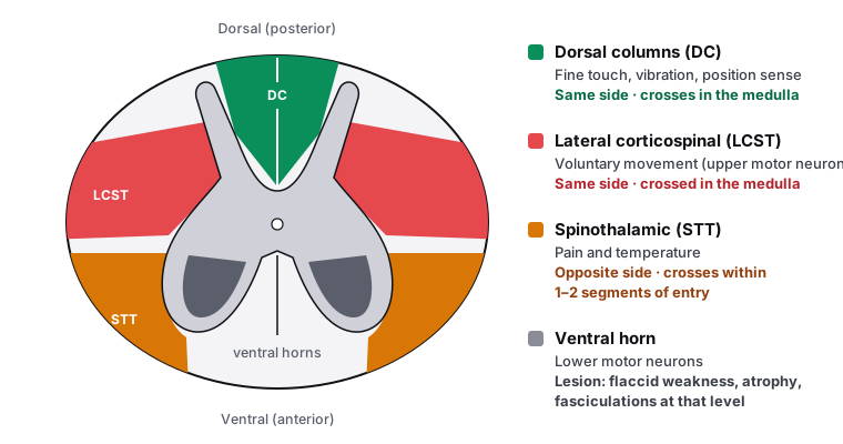
A lesion of the corticospinal tract gives upper motor neuron signs below it: spastic weakness, hyperreflexia and an upgoing (Babinski) plantar response. A lesion of the ventral horn gives lower motor neuron signs at that level: flaccid weakness, atrophy, fasciculations and lost reflexes.
Six lesions, one picture each
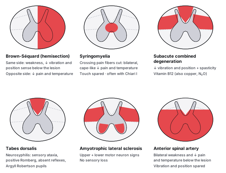
| Syndrome | Typical cause | Tell-tale clue |
|---|---|---|
| Brown-Séquard | Penetrating trauma (stab wound) | Pain loss on one side, weakness on the other |
| Syringomyelia | Chiari I malformation, trauma | Pain and temperature loss in both arms ("cape"), painless burns on the hands; ventral horn and Horner signs as the syrinx grows |
| Subacute combined degeneration | Vitamin B12 deficiency (also copper deficiency, nitrous oxide) | Vibration loss plus spasticity and Babinski signs; macrocytic anemia |
| Tabes dorsalis | Tertiary syphilis | Wide-based gait, positive Romberg, Argyll Robertson pupils, Charcot joints |
| ALS | Degeneration of upper and lower motor neurons | Fasciculations next to hyperreflexia; sensation and bladder spared |
| Anterior spinal artery infarct | Aortic aneurysm repair or dissection, hypotension | Sudden paraplegia with vibration and position sense intact |
High-yield details by syndrome
- Brown-Séquard: at the level of the lesion, all sensation is lost on the same side, with lower motor neuron signs. A lesion above T1 can add an ipsilateral Horner syndrome.
- Syringomyelia: usually starts around C8–T1. As it expands into the ventral horns, the intrinsic hand muscles waste. Diagnose with MRI.
- Subacute combined degeneration: can appear before (or without) anemia. Methylmalonic acid is raised. Nitrous oxide inactivates B12, so it can unmask deficiency after anesthesia or recreational use. Late damage may not reverse with B12.
- Tabes dorsalis: damages the dorsal roots as well as the columns: shooting "lightning" pains, absent reflexes, Charcot joints. Test the CSF (VDRL); treat with IV penicillin G.
- ALS: spares eye movements and bladder control. Some familial cases have SOD1 mutations. Riluzole (reduces glutamate release) modestly prolongs survival.
- Anterior spinal artery: the lower cord depends on the artery of Adamkiewicz, a large feeder usually arising between T9 and T12, which is why aortic surgery and dissection threaten it.
- Pure lower motor neuron disease (ventral horns only): poliomyelitis, and spinal muscular atrophy, where type 1 (Werdnig-Hoffmann) is a floppy infant with tongue fasciculations from SMN1 mutations.
Upper or lower motor neuron?
| Sign | Upper motor neuron | Lower motor neuron |
|---|---|---|
| Tone | ↑ (spastic) | ↓ (flaccid) |
| Reflexes | ↑, with clonus | ↓ or absent |
| Babinski sign | Present | Absent |
| Atrophy | Mild (disuse) | Marked |
| Fasciculations | Absent | Present |
Right after an acute cord injury, spinal shock can make an upper motor neuron lesion look flaccid and areflexic for days to weeks before spasticity appears.
Three more you will see
| Syndrome | Typical setting | Findings |
|---|---|---|
| Central cord | Neck hyperextension in an older adult with cervical spondylosis | Arms (especially hands) weaker than legs; variable sensory loss |
| Conus medullaris | Lesion at about the L1–L2 vertebral level | Sudden, symmetric saddle anesthesia; early bladder and bowel dysfunction; can mix upper and lower motor neuron signs |
| Cauda equina | Large central disk, tumor, epidural abscess | Asymmetric radicular pain, lower motor neuron signs only, saddle anesthesia, urinary retention. Emergency MRI and decompression |
Reflexes and their roots
| Reflex | Root |
|---|---|
| Biceps | C5–C6 |
| Brachioradialis | C6 |
| Triceps | C7 |
| Patellar | L4 (L3–L4) |
| Achilles | S1 |
| Cremasteric · anal wink | L1–L2 · S3–S4 |
Practice questions
Question 1NeurologyBrown-Séquard syndrome
A 24-year-old man is stabbed in the back. Examination shows weakness and loss of vibration sense in the right leg, and loss of pain and temperature sensation in the left leg. Where is the lesion?
A left-sided hemisection would cause weakness and vibration loss in the left leg, with pain and temperature loss on the right. This patient has the opposite pattern: the same-side motor and vibration deficits mark the side of the lesion.
Corticospinal and dorsal column fibers have already crossed in the medulla, so a hemisection causes weakness and vibration loss on the same side as the lesion (right). Spinothalamic fibers cross within 1–2 segments of entering the cord, so pain and temperature are lost on the opposite side (left), starting 1–2 levels below the lesion. This is Brown-Séquard syndrome.
Bilateral dorsal column damage would cause loss of vibration and position sense in both legs, with no weakness and normal pain and temperature. It is the pattern of tabes dorsalis or vitamin B12 deficiency.
Anterior two-thirds (anterior spinal artery) infarction causes bilateral weakness and bilateral loss of pain and temperature, with vibration and position sense spared. Here the findings are asymmetric and vibration is lost.
A central cord lesion (syringomyelia, central cord syndrome) damages the crossing spinothalamic fibers: bilateral loss of pain and temperature at that level, often in a cape-like distribution, with vibration and position sense spared. It does not cause one-sided weakness with vibration loss.
The answer is B.
Key takeaway
In a cord hemisection, weakness and vibration loss are on the same side as the lesion, and pain and temperature loss is on the opposite side.
Brown-Séquard syndrome is a hemisection of the spinal cord, classically from penetrating trauma. The corticospinal tract and dorsal columns cross in the medulla, so they are damaged on the same side as the lesion. The spinothalamic tract crosses within a segment or two of entering the cord, so its loss appears on the opposite side.
- Same side: weakness (upper motor neuron signs), loss of vibration and position sense
- Opposite side: loss of pain and temperature, beginning 1–2 segments below the lesion
- At the level of the lesion: loss of all sensation on the same side, with lower motor neuron signs
- Common causes: stab and gunshot wounds, tumors
Question 2NeurologySubacute combined degeneration
A 58-year-old woman who follows a strict vegan diet has numb feet and unsteady walking for 6 months. She has lost vibration and position sense in both legs, her legs are spastic and both plantar responses are upgoing. Her hemoglobin is low and her MCV is 112 fL. Which structures are damaged?
Ventral horn damage (polio, spinal muscular atrophy, ALS) causes lower motor neuron signs: flaccid weakness, wasting and fasciculations. This patient is spastic with upgoing plantar responses and has sensory loss.
Dorsal column damage alone (as in tabes dorsalis) explains the loss of vibration and position sense and a sensory ataxia, but not the spasticity and Babinski signs. Those need corticospinal involvement.
Vitamin B12 deficiency causes subacute combined degeneration. The loss of vibration and position sense is the dorsal columns; the spasticity, hyperreflexia and upgoing plantar responses are the lateral corticospinal tracts. A strict vegan diet and a macrocytic anemia (MCV 112 fL) complete the picture.
Spinothalamic damage would reduce pain and temperature, which are typically spared in B12 deficiency. Ventral horn damage would give flaccid weakness, not spasticity.
The anterior white commissure carries crossing spinothalamic fibers. Damage there (syringomyelia) causes a cape-like loss of pain and temperature with vibration and position sense preserved: the opposite of this patient.
The answer is C.
Key takeaway
B12 deficiency damages the dorsal columns and lateral corticospinal tracts: loss of vibration and position sense plus spasticity, with a macrocytic anemia.
Vitamin B12 is needed for myelin synthesis. Without it, demyelination develops in the dorsal columns and lateral corticospinal tracts, hence the name subacute combined degeneration. Strict vegans, patients with pernicious anemia or ileal disease, and long-term metformin or nitrous oxide users are at risk. Neurologic damage can appear before the anemia, and late damage may not reverse with treatment.
- Dorsal columns: loss of vibration and position sense, sensory ataxia, positive Romberg sign
- Lateral corticospinal tracts: spasticity, hyperreflexia, upgoing plantar responses
- Macrocytic anemia with hypersegmented neutrophils; raised methylmalonic acid and homocysteine
- Treat with B12: folate alone can correct the anemia while the nerve damage progresses
Question 3NeurologyAnterior spinal artery infarction
A 71-year-old man has open repair of an abdominal aortic aneurysm. When he wakes, he cannot move his legs and cannot feel a pinprick below the umbilicus. He can feel a tuning fork on both big toes and knows which way they are moved. What is the most likely diagnosis?
The anterior spinal artery supplies the anterior two thirds of the cord, including the corticospinal and spinothalamic tracts, so infarction causes bilateral weakness and loss of pain and temperature below the lesion. The dorsal columns, supplied by the posterior spinal arteries, are spared, so vibration and position sense are intact. Aortic surgery can compromise the artery of Adamkiewicz, which feeds the lower anterior spinal artery.
Brown-Séquard syndrome is an asymmetric hemisection: one-sided weakness and vibration loss with opposite-sided pain loss. This patient's deficits are bilateral and his vibration sense is spared.
Subacute combined degeneration develops over weeks to months and damages the dorsal columns (vibration is lost) and the corticospinal tracts. It does not appear suddenly after surgery and it does not spare vibration.
Syringomyelia develops slowly and causes a cape-like loss of pain and temperature in the arms with preserved touch, often with a Chiari I malformation. It does not cause sudden paraplegia.
Tabes dorsalis (tertiary syphilis) damages the dorsal columns, causing loss of vibration and position sense, sensory ataxia and lightning pains. This patient's vibration and position sense are intact.
The answer is A.
Key takeaway
Sudden paraplegia with loss of pain and temperature but preserved vibration and position sense, after aortic surgery or dissection, is anterior spinal artery infarction.
The cord has one anterior and two posterior spinal arteries. The single anterior artery supplies the anterior two thirds, so its occlusion damages the motor and pain and temperature pathways on both sides while sparing the dorsal columns. The lower cord depends heavily on the artery of Adamkiewicz, which makes aortic surgery and aortic dissection the classic triggers.
- Bilateral weakness and loss of pain and temperature below the lesion
- Vibration, position sense and light touch are preserved (dorsal columns)
- Causes: aortic aneurysm repair, aortic dissection, severe hypotension
- Spinal shock (flaccid, areflexic) comes first; spasticity develops later
Go deeper in the Knowledge Hub: spinal cord tracts and reflexes, incomplete spinal cord syndromes, syringomyelia and ALS.